Electrical impulses course through our heart and keep it beating. That's why a jolt from an automated external defibrillator can boost it back into action if the beating stops. But new research says there may be more to keeping a heart beating than just electrical impulses.
The new research, published online in the journal Cell, reports that a type of white blood cell called a macrophage is abundant in healthy heart muscle tissue and — in a new role for the cells — contribute to the mechanism that makes our heart beat.
Macrophages can be found in most tissues and are best known for their role in the immune system. They find foreign particles (including bacteria and viruses), process them, and present them to other cells of the immune system so that a response may be mounted. Macrophages also engulf and digest foreign materials in a process called phagocytosis, and release some chemicals that instruct other cells in the immune response.

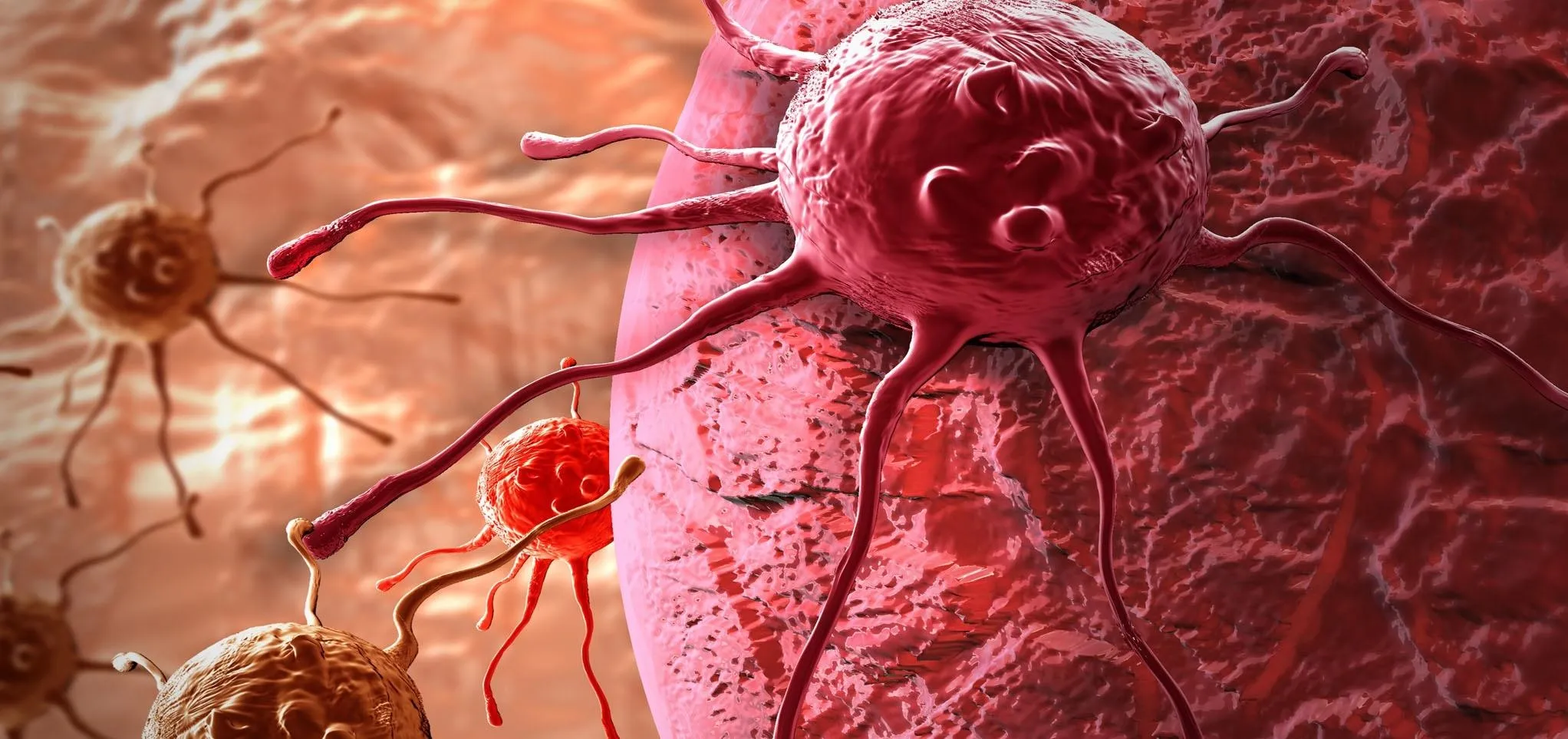
Macrophages with projectile-looking surfaces that are interacting with round lymphocytes
We know macrophages play a role in repairing damage to the heart by removing cell debris and assisting in remodeling (i.e., healing) the heart cells, but Dr. Matthias Nahrendorf of Harvard Medical School in Boston, along with his colleagues, wanted to know why they found macrophages in healthy heart muscle.
The scientists found that macrophages are abundant in the atrioventricular node of human and mouse heart tissue. That node, a group of cells, is a part of the system that conducts electrical impulses through the ventricles of the heart — the part of the heart that pumps blood out of the heart and through the body.
Experiments on mice showed that macrophages express a protein called connexin 34 and connect to heart muscle cells via gap junctions — pore-like structures that function to coordinate heart muscle contractions. Mice lacking a key gap junction protein showed an abnormal slowing of the electrical signal through the atrioventricular node. When tissue macrophages are depleted in the mice, the mice develop a complete block of the atrioventricular signal.
This type of atrioventricular block occurs in humans and is treated with a pacemaker to maintain a healthy heart beat rhythm.
The new study clearly showed that macrophages play a heretofore unknown and critical role in maintaining the normal beat of the heart.
Conducting the Heart's Beat
The cardiac conduction system is the orchestrator of the heart's electrical impulses — real electrical signals that cause the heart to contract so that blood is moved through and out of it.
An electrical impulse begins in the sinoatrial node, a collection of nerves and tissue in the upper right atrium of the heart. From there, the electrical impulse flows through to activate both atria, the upper chambers of the heart that hold blood. The impulse causes them to contract.
The signal also moves to the ventricles (the lower chambers of the heart) through the atrioventricular node, the bundle of His, and the Purkinje fibers. The signal slows down as it passes through the atrioventricular node, a slowing that allows the ventricles enough time to finish filling with blood before they contract to move the blood out of the ventricles to the lungs and rest of the body.
The new research by Nahrendorf and colleagues puts macrophages prominently in this scenario. They reside in normal heart tissue and assist in conducting electrical impulses through the atrioventricular node.
The study authors suggest macrophages in the heart may be a new target for therapeutic intervention of abnormal heart rhythms. It's a long way from scavenging foreign particles and a very new role for macrophages, indeed.
- Follow Invisiverse on Facebook and Twitter
- Follow WonderHowTo on Facebook, Twitter, Pinterest, and Google+
Cover image via PublicDomainPictures/Pixabay












Comments
Be the first, drop a comment!