
Using extreme time-lapse microscopy, scientists watched a virus take over a bacteria to create a cell that looked and functioned more like a plant or animal cell. True story.
In a study published in Science, researchers from the Division of Biological Sciences and Department of Chemistry and Biochemistry at the University of California, San Diego, as well as the Department of Biochemistry and Biophysics at the University of California, San Fransisco, took a closer look at how microbes replicate themselves by hijacking the cell of another microbe. While considered standard operating procedure in the microbe world, this study looked specifically at a very cool and common type of virus called a bacteriophage.
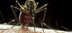
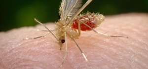
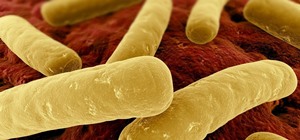
Usually called a phage, a bacteriophage is a virus with a big appetite for bacteria. With a name that roughly means bacteria-eater, phages are a diverse and interestingly shaped group of viruses that show up wherever bacteria are, including your lunch and, correspondingly, your gut.
Bacteriophage are the subject of intense research as medical science races to find non-drug alternatives to fight drug-resistant bacteria. Antibiotics are continuing to lose effectiveness due to overuse and the exceptional adaptive capabilities of bacteria. But specially designed viruses may be able to fight off infections with the bacteria developing resistance, since the virus can evolve right along with it.
In the National Action Plan for Combating Antibiotic-Resistant Bacteria, the White House lists the use of phage and phage-derived processes to target specific bacteria without harming human microbiota.
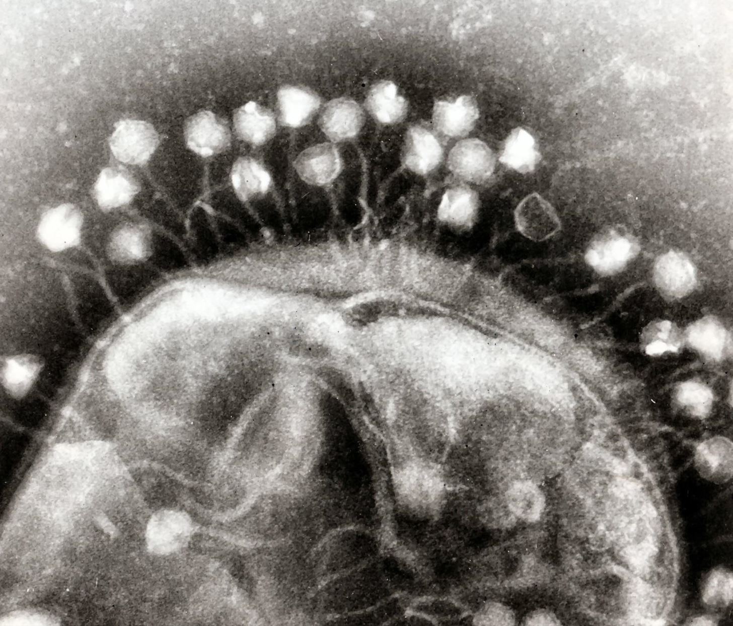
Using florescent microscopy and cryo-electron tomography, researchers in the Science study logged the action of bacteriophages as they attached to Pseudomonas chlororaphis, a soil-dwelling bacteria that is itself used to deter fungal infections in agricultural crops like cucumbers and peppers.
Florescent microscopy is often used to see and create images of microorganisms. These microscopes utilize intense light sources, like lasers, to illuminate proteins tagged with a florescent compound. Another process invaluable to viewing microorganisms as small as a virus is cryo-electron tomography. This process offers higher resolution images and data and is frequently combined with florescent microscopy.
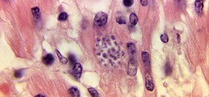
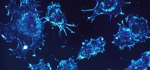
In this study, scientists used these technologies to observe phages as they took over the cellular function of P. chlororaphis. Researchers were able to document the path taken by the virus to create a surprisingly complex structure beyond what either the bacteria, or the virus, were able to make before infection. Take a look at the proteins moving around the virus and bacteria in the short video below:
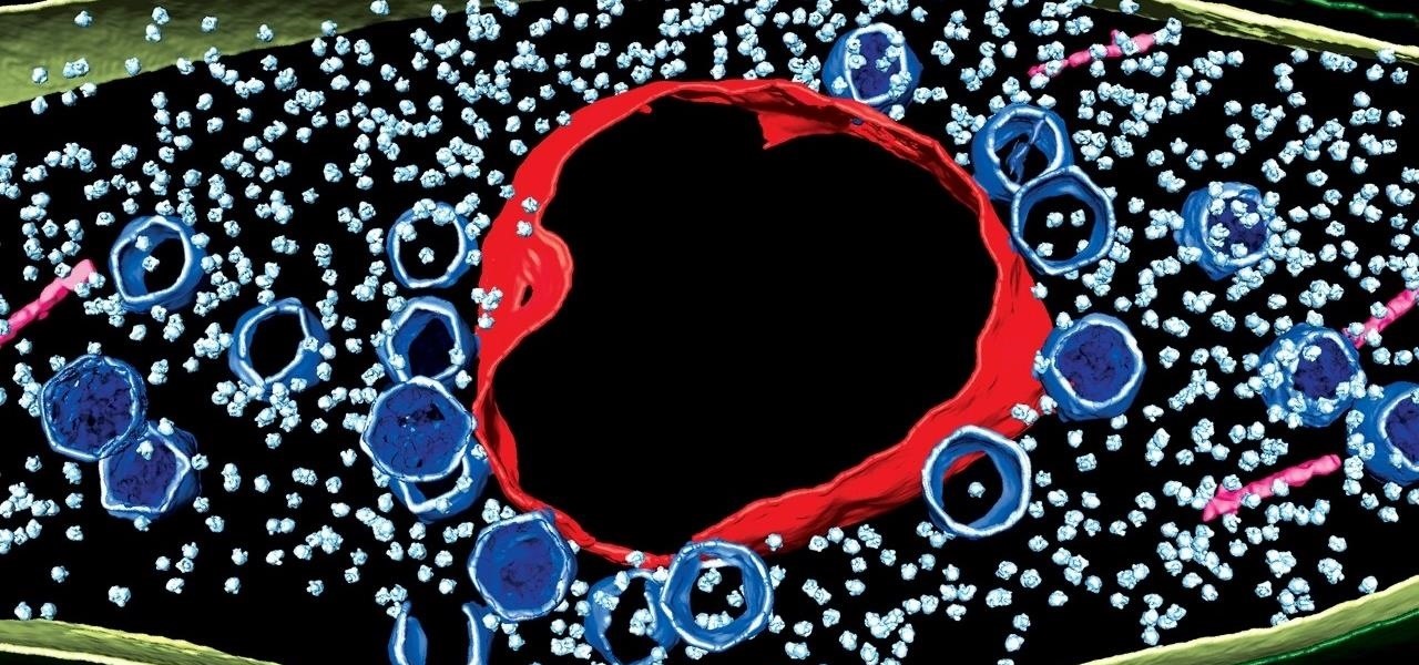
What's really fascinating is that the virus and bacteria come together to make something only seen in plant and animal cells—a structure that looks like a nucleus.
Fables of the Bacterial Reconstruction
The bacteriophage infiltrates the bacteria cell, dissolving most of its internal structure with genetic material injected by the phage. Using the remaining structure, the virus rebuilds the cell to replicate, generating a nucleus-like compartment (shown in red in this article's cover image, and shown in blue in the video clip). What is different about this? Neither viruses or bacteria usually have an enclosed nucleus.
"Scientists have been studying viruses for a hundred years, but we've never seen anything like this before," study researcher Joe Pogliano, a professor of molecular biology at UC San Diego, said in a press release. "This factory and the surrounding arrangement of the infected cell are remarkably similar to the organization seen in plant and animal cells."
The end result of this cellular reconstruction occurs when the cell bursts, spilling out viral material that, in this case, will infect surrounding bacteria.
The findings that a virus can instruct a bacteria to create a structure more complex than either—complete with cellular compartments that enclose DNA processes and exude proteins outside that compartment—point to abilities usually displayed by eukaryotic cells, the type that make up plants and animals.
Eukaryota are one of the three domains of life, alongside Bacteria and Archaea. Established in the 1970s, these domains form a classification level more basic than kingdoms. None of these domains are currently thought to possess an ancestor—they're the oldest and biggest groups we've found. Animals, plants, and fungi are classified as eukaryotes.
Viral eukaryogenesis is a theory that suggests multicellular (eukaryote) organisms evolved from virus cells that infected bacteria to create a more compound critter—the eukaryote, and eventually plants, animals, and us. As shown in this research, viral infection of bacteria does create more sophisticated machinery—the enclosed nucleus that resembles a mammalian cell organelle.
It may be too early to know if this particular virus is an intermediate step in the transition from bacteria and viruses to multicellular eukaryotes, but this discovery could broaden knowledge about the origins of life as we know it.
Part of our ancient past, or a potential life-saving antibacterial option of the future? Bacteriophages may have it all.
Just updated your iPhone? You'll find new emoji, enhanced security, podcast transcripts, Apple Cash virtual numbers, and other useful features. There are even new additions hidden within Safari. Find out what's new and changed on your iPhone with the iOS 17.4 update.


Be the First to Comment
Share Your Thoughts